 |
 |
|
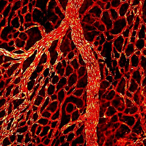
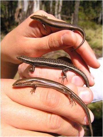
| Uterine vasculature of the viviparous skink, Eulamprus quoyii (confocal image) | Uterine epithelium of the oviparous skink, Ctenotus taeniolatus (scanning electron micrograph) | Viviparous Australian skinks with complex placentation: Pseudemoia entrecasteauxii (top), P. spenceri (middle), P. pagenstecheri (bottom) |
Evolution of viviparity and placental complexity in squamate reptiles
The evolutionary transition from oviparity (egg-laying) to viviparity (live-bearing) is one
of the most fundamental, yet least understood processes in evolutionary physiology.
The evolution of viviparity involves modifications to a suite of integrated physiological
features whereby the uterus and extra-embryonic membranes are transformed from
functionally simple structures in oviparous species to complex structures in which
respiratory gases, water, and nutrients are transported between mother and embryo in
viviparous species. My research focuses on squamate reptiles (lizards and snakes)
because they are the best animal model for studying the evolution of viviparity, with at
least 108 independent origins of viviparity within the group.
Three major events are required for viviparity to evolve from oviparity: 1)
prolonged retention of embryos in the oviduct, 2) thinning and/or loss of the eggshell, and 3) the
elaboration of the placental vascular bed to facilitate increased transport of oxygen and nutrients
to accommodate the metabolic demands of the growing embryo. My current research focuses
primarily on the third of these events because in utero oxygen availability appears to be one of
the primary factors limiting the amount of development attained by embryos during gestation.
During my post-doctoral research with Mike Thompson and Chris Murphy at the University of Sydney, I used laser scannning confocal microscopy obtain detailed three dimensional images of placental blood vessels throughout pregnancy in several species of oviparous and vivparous Australian skinks. My current research builds on these previous studies by determining which key molecular factors control placental angiogenesis in squamate reptiles and how expression of these factors is modified during the evolution of viviparity. For my current research I use immunohistochemistry and light microscopy to identify presence and distribution of the important angiogenic factors vascular endothelial growth factor (VEGF) and hypoxia-inducible factors (HIFs) during the reproductive cycle, and how expression of these factors correlates with blood vessel proliferation. Because this project relys heavily on the use of histological techniques, the work may be of particular interest to students interested in animal physiology as well as medical sciences.